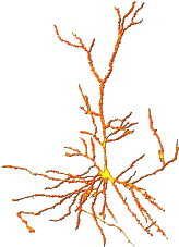
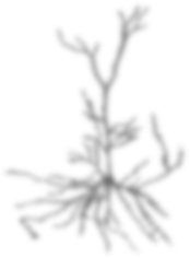
Image of the Day
The images that appear in the "Image of the Day" are selected for the freshness of their views on Brain Mapping, their esthetic appeal, their quirkiness, or someimes just to prod you into thinking about the field and its context. Their appearance here is not an endorsement of their subject matter.
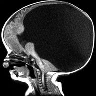 |
Holoprosencephaly Neuroimaging
NEUROIMAGING EVALUATION of HPE is best accomplished by MRI. In all suspected cases the scan should include axial, sagittal and coronal sequences to enable complete evaluation of midline structures. Also, thin sections are recommended; ideally no greater than 3 mm thick, with spacing of 0.5 mm. Even better are volumetric protocols, which generally have 1.5 mm slice thickness with no gaps.Until now, a lack of standardization of protocols has made it difficult to meaningfully classify this disorder. The individuals to the right have been instrumental in helping the Carter Centers to develop standardized MRI sequences to obtain 3-dimensional structural data for HPE, which can then be compared across many institutions. It is hoped that utilization of improving technology to fully characterize brain structure in HPE will lead to better classification schemes, important genotype-phenotype correlations, and ultimately to a better understanding of etiologies. |
For more information on this image, visit:
[ This Link ]
Submitted by:
Mark Cohen
|