|
|
The Images of the Day
 | Your brain on shoppingHow can my husband and I have such different approaches to the shopping experience? I attribute it to wiring in the brain, and I think it applies to many men and women out there.As a refresher, remember that the left brain is the logical, analytical side of the brain - it is the worker bee, focusing and analyzing one thing at a time. The right brain is free to play - its the home of imagination, emotional memory and bonding with others. With four times as many connections between the left and right hemispheres of the brain, women are four times as likely to tap into the right brain when encountering a situation. Many factors come into play when going through the shopping process for women - its all about the big picture.
Click here for more information | 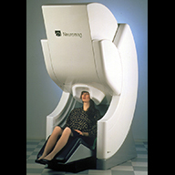 | MEG in OperationThe Neuromag VectorView MEG instrument from Elekta.
Click here for more information | 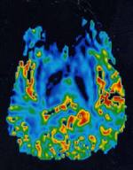 | Blood flow mapping in epilepsyBlood flow often increases in brain tumors. MRI perfusion studies can demonstrate this phenomenon. This Figure shows the increased blood flow.
From Unviersity of Washington.
Click here for more information | 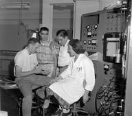 | Brain Mapping at LBLHilac Brain Mapping experiment.Members of the Tobias group check the beam intensity for a "brain mapping" experiment. (L. to r.) John Lyman, Howard Chung, Nicholas Yanni, Jean Luce, In the experiment, the researchers were able to make lab animals blink by directing a beam of invisible alpha radiation into the cornea of their eyes. This was one of several experiments underway in 1961 using particle beams to explore the brain.
Click here for more information | 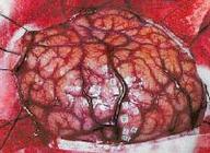 | Epilepsy SurgeryThe surface of the brain of a patient undergoing epilepsy surgery evaluation. The surgeon has electrically stimulated different brain locations and labeled each location with a numbered tag. The responses to electrical stimulation may include motor movements, sensory responses and, in some cases, electrically induced partial seizures (afterdischarge). These responses are recorded and used for planning placement of a subdural electrode array and later surgery.
Click here for more information |  | The University of Washington Human Brain ProjectThe UW Human Brain Project is one project within the national Human Brain Project, a national multi-agency effort to develop informatics tools for managing the exploding amount of information that is accumulating about the human brain.The objective of the UW Human Brain Project effort is to organize functional information about the brain around the structural information framework that is the long term goal of our work. This application therefore extends the utility of the Digital Anatomist Project by using it to organize non-structural information
Click here for more information | 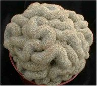 | Brain CactusMammillaria elongata cristata, known as the "Brain Cactus", forms attractive undulating fan-like stems covered in dense golden spines. Cristate forms generally occur when injury occurs to the plant at a young age (this damage can be due to insects eating the growing tip, or from many other causes, including a genetic predisposition). In reaction to the "injury", the cells at the tip of the branch where growth occurs begin to multiply at a much faster rate and the normal growing tip "goes crazy", creating fantastic whorls and fans. Mammillaria elongata, in habitat, lives in crags between rocky outcroppings in Central Mexico, eventually forming long cascades of stems with pendant offsets. In this habitat, the water drains quickly away from the roots of the plant, never allowing the plant to remain waterlogged. For this reason, it is essential in cultivation to use a very porous soil, which will allow quick drainage. Bright, filtered light. Water thoroughly when soil is dry to the touch. As the "fans" of cristates can trap water that can lead to rot, it is preferable to water from beneath, if possible. Water thoroughly when soil is dry to the touch, except on cloudy, humid or cold winter days. Protect from frost. Shipped established in a 3.5" pot.
Click here for more information | 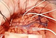 | Epilepsy Surgery and Brain Mapping ProgramThe Epilepsy Surgery and Brain Mapping Program at The University of Mississippi Medical Center is a comprehensive service offering diagnosis and management of adult and pediatric patients with intractable epilepsy. The multidisciplinary team is comprised of experts in Epileptology, Adult and Pediatric Neurosurgery, Neurology, Neuropsychology, Neuropharmacology and Neurophysiology. Patients with medically intractable seizures or with structural lesions involving eloquent brain regions may be considered for invasive monitoring and surgical intervention.
Click here for more information |  | Landscape of the MindUCLA has constructed the worlds most comprehensive atlas of the human brain.
International Consortium for Brain Mapping
National Institutes of Health: $5.5MAs many ways as our brains are similar, there are subtle differences that make each unique. Consequently, it is difficult to know what within the human brain is normal and what is not. Neurologists may be well acquainted with the brains gross anatomy, but this absence of exactitude poses problems that have potential implications for researchers as well as for surgeons trying to navigate the corrugations of this most essential organ. Now there is the worlds most comprehensive atlas of the brain, developed by a multidisciplinary team of UCLA researchers with colleagues in six other countries and at two U.S. universities. The atlas database comprises high-definition structural maps from gross anatomy to microscopic detail based on scans of the brains of some 7,000 individuals and referenced for age, race, gender, educational background, genetic composition and other distinguishing characteristics. Layered over the anatomical maps are brain functions such as memory, emotion, language and speech. The need for such a tool is evident; within the past 12 months there have been more than 1.5 million visits to the consortiums Web site and 43,000 downloads of highly detailed information. And the database in some respects the equivalent of the human genome project continues to grow as researchers plug in new information.
Click here for more information | 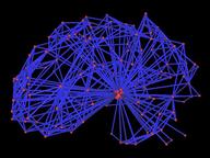 | Visual ComplexityAs an undergraduate at UC Davis, Patrick Yau had the opportunity to work at the Visualization and Graphics Research Group in the Institute for Data Analysis and Visualization (IDAV). He was under the guidance of Professor Bernd Hamann and Dr. Lars Linsen when he and his colleagues developed the project "Brain Mapping via Hierarchical Isosurface Segmentation Based on Discrete Curvature".The idea of the project is to look into the surface of two brains and identify the similarities between then. They focused in particular on the curvature of the brains (folds and bumps). Yau worked on data preprocessing and the automated brain mapping algorithm. He wrote a procedure that automatically identifies the front, side and the top for two brains using Principal Component Analysis (PCA), and he also wrote a procedure that constructs a topology graph based on the curvature of a brain. The two images shown here reveal different representations of the same brain: as points and as a topology graph.
Click here for more information | 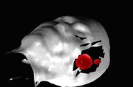 | NASA Infrared Camera Helps Surgeons Map Brain TumorsUsing an infrared video camera developed by researchers at NASA's Jet Propulsion Laboratory in Pasadena, Calif., surgeons are testing thermal imaging and image processing to see if they can create useful maps of brain tumors.Researchers want to see if the camera, which detects infrared -- or heat -- emissions might help neurosurgeons better visualize tumors before they operate and also find tiny clusters of cancerous cells that might remain after surgery.
Click here for more information | 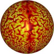 | New Institute to Seeks to Serve Physicists and EngineersMedical physicists and bioengineers aim to gain visibility, funding, and clout with the creation of the National Institute of Biomedical Imaging and Bioengineering (NIBIB), which became the latest addition to the National Institutes of Health family when it was signed into law in the waning hours of the Clinton presidency.Mapping the BrainNIBIB was born of frustration: "People who work in engineering and imaging have major difficulties selling their projects to NIH," says Ferenc Jolesz. He should know. As head of magnetic resonance imaging and image-guided therapy programs at Harvard Medical School's Brigham and Women's Hospital, Jolesz oversees about 100 researchers and an annual budget of $10 million. "We have physicians, engineers, mathematicians, physicists, computer scientists. Our projects can be good for many things--the brain, the heart, the lungs. It's very difficult to fit into the culture of NIH, which is driven by organ- or disease-oriented research. You have to fake it and say you are doing something for a specific disorder."
Click here for more information | 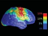 | Daring To Take Risks and Reap the RewardsAt the University of California, Los Angeles, Paul Thompson and his colleagues developed a novel computational framework that effectively stretches, contorts, and changes the geometry of highly image of Innovative brain-mapping techniquesdetailed three-dimensional brain images obtained via magnetic resonance imaging (MRI). These manipulations allow scientists to overlap and meld multiple brain images, collected over time or from multiple individuals, and enable comparisons between normal and dysfunctional brains. To date, the images have clearly revealed the changes wrought by Alzheimer's disease, methamphetamine abuse, schizophrenia, and AIDS. "With R21 funding, we developed new mathematical methods for understanding the effects of disease," says Thompson, an associate professor of neurology. "These images are really snapshots of a disease spreading over time."
Click here for more information |  | Your Brain's Rooting for You But Do You Benefit?Brain-based Business Insights.
Click here for more information | 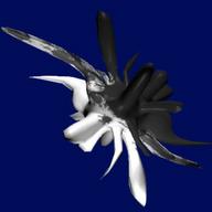 | The Einstein's Brain ProjectFor the past 8 years Alan Dunning, Dr. Morley Hollenberg and Paul Woodrow, of the art and science collaboration, "the Einstein's Brain Project," have been investigating ideas about consciousness through immersive and mixed virtual reality. The project has brought together artists, scientists, technologists and others in a transdisciplinary space tp develop new ways of representing and understanding the technologized body.The shapes of thought is a work that visualizes EEG and other bioelectrical signals as three-dimensional forms. The forms are generated by monitoring the EEG of a participant recalling a traumatic event and using the numbers to change simple primitives to complex meshes. Each vertext on a primitive is assigned a point in space and each is pushed and pulled by the incoming EEG data. Over very long periods of time - more than 12 hourse in some cases - a smooth sphere or cube becomes a heavily fissured, bumpy and spiked object - a recent geological record of the EEG pattern generated by the participant. At prearranged intervals, the form is saved into a database to allow the event path to be retraced in the future.
Click here for more information | 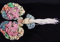 | Karen Norberg - Knitted Brainthe Museum of Scientifically Accurate Fabric Brain ArtTo neuroscience researchers, the human brain is a complex organ riddled with mystery. But to developmental psychologist Marjorie Taylor and psychiatrist Karen Norberg, it's also an inspiration for unique quilts, knitting, and other work that they showcase in an online museum of "scientifically accurate fabric brain art."The two were drawn to the niche independently. Taylor, a professor at the University of Oregon, Eugene, had been making quilts on the side for years before she turned her needle to neuroscience. Struck by the cover images of journals like Cerebral Cortex, she began reproducing them in fabric, creating pieces that--for example--show positron emission tomography scans of the brain's response to hearing or seeing words. Karen Norberg who works at the National Bureau of Economic Research in Cambridge, Massachusetts, says she began knitting a brain to kill time when she was undergoing clinical training in child psychiatry. The product now resides at the Boston Museum of Science. This work was profiled at the AAAS/Science web site
Click here for more information | 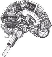 | Left Brain - Don StewartDon Stewart received his BS degree in Biology and Art at Birmingham-Southern College in 1981, paying his way with a nightclub act as a comic ventriloquist. Initially he enrolled in art classes as a respite from the rigors of premedical studies. Soon, however, he was exposed to the process of composite imagery, and began exploring techniques in ballpoint drawing. Don continued to pursue artistic interests as a hobby during medical training at the University of Alabama at Birmingham.After receiving his M.D. in 1985, Dr. Stewart moved north to Minnesota, to complete an internship in surgery at the Mayo Clinic. The lifestyle of a surgical resident proved, however, to be at odds with his creative side. By year's end, Don opted to discontinue his medical studies, after accepting awards for both short fiction and poetry, and publishing his first two composite drawings. He has since worked as a graphic artist, writer, and creative consultant through his company, DS Art.
Click here for more information |  | Pre-Colombian Brain SurgeryRock art, and healed trephined skulls, from the early Peruvians show considerable sophistication in performing open brain surgeries. It is not clear why such surgeries were performed. The link below presents some of this ample history.
Click here for more information | 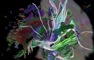 | Tensorlines and superquadric glyphsGordon Kindlmann, Dr. David Weinstein, Scientific Computing and Imaging Institute, University of UtahSome tensor-line fiber tracts and superquadric tensor glyphs used to depict some of the white matter structure in a DT-MRI scan. The tensorlines have been highlighted for emphasis. Determining the extent to which the the computed fiber tracts actually correspond to white matter pathways is a subject of ongoing work.
Click here for more information |  | High Resolution fMRI of the Cat at 4TfMRI in a cat at 9.4 Tesla. The color is high resolution (.15x.15x2 mm) BOLD activation patterns in response to visual stimulation across the LAYERS in primary visual cortex, overlayed on a high resolution T1 image.Noam Harel, Joseph Lin, Steen Moeller, Kamil Ugurbil, and Essa Yacoub "Combined imaging.histological study of cortical laminar specificity of fMRI signals"
Click here for more information |  | a case of phrenology - finalBy marcyintellect
Click here for more information | 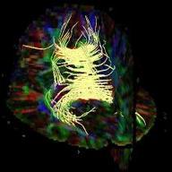 | Diffusion Imaging, Diffusion Tensor Imaging and Fiber TrackingRecent advances in magnetic resonance research have opened up new opportunities for gathering functional information about the brain. The development of diffusion imaging and diffusion tensor imaging (DTI) has offered the possibility to go beyond anatomical imaging and study tissue structure at a microscopic level in vivo. Currently, DTI provides an invaluable tool for the study and diagnosis of white-matter diseases.
Increasing research focus has been placed on studying neuronal connectivity. An important application of DTI is fiber tracking, which allows elucidation of white-matter tracts. Combined with fMRI, information about white-matter tracts reveals important information about neurocognitive networks, and may improve our understanding of brain function. Recently, the potential of applying SENSE to DTI was demonstrated at 3.0 Tesla to improve acquisition speed and image quality. These studies have demonstrated the feasibility of fast fiber tracking in the human brain. Future research will concentrate on improving the accuracy and robustness of the technique, as well as enhanced visualization of the white fiber tracts in three dimensions.dipl. phys. Thomas Järmann, dipl. inf. ETH Philipp Stämpfli, Dr. Klaas Prüssmann, Prof. Dr. Peter Boesiger
Click here for more information | 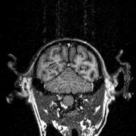 | Stereotaxic MRI Brain Atlas of the MonkeyThese images were taken at the Laboratory for Magnetic Resonance, Imaging and Spectroscopy, National Institutes for Physiological Sciences, Myodaiji, Okazaki 444, Japan. This work was supported by the joint research program of National Institute of Physiological Sciences.Masato Taira Associate Professor
Department of Physiology
Nihon University, School of Medicine
TEL: +81-3-3972-8111 ext.2231
FAX: +81-3-3972-8292
masato@med.nihon-u.ac.jp
Click here for more information | 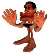 | The Sensorimotor HomunculusThe representation of the body onto the brain preserves general connectedness and topology, but the amount of cortical surface dedicated to body regions is scaled such that some muscle groups and skin regions project to or from larger brain areas than do others.
Click here for more information | 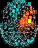 | Java EEG AnalyserThis program reads and displays electro-encephalogram (EEG) data produced by the device manufactured by Electrical Geodesics Inc. It interactively analysis the dimension of the data and finds convincing linear generators for the EEG data. Thanks to Thomas Gruber at Liverpool University for the data and the idea.The latest release, containing all the source code (protected by the GNU General Public License), example files and this html documentation can be downloaded from sourceforge/projects/jeeg You can use the source code to adapt this program to work with any other device with a different file format, as well as overcome the limited user interface.
Click here for more information |  | Electronic Chip Interacting with the BrainScientists at the University of Washington (UW) are working on an implantable electronic chip that may help establish new nerve connections in the part of the brain that controls movement. Their most recent study, would be reported in the Nov. 2, 2006, edition of Nature, showed such a device can induce brain changes in monkeys lasting more than a week. Strengthening of weak connections through this mechanism may have potential in the rehabilitation of patients with brain injuries, stroke, or paralysis.The authors of study, titled "Long-Term Motor Cortex Plasticity Induced by an Electronic Neural Implant," were Dr. Andrew Jackson, senior research fellow in physiology and biophysics, Dr. Jaideep Mavoori, who recently earned a Ph.D. in electrical engineering from the UW, and Dr. Eberhard Fetz, professor of physiology and biophysics. For a number of years Fetz and colleagues have studied how the brains of monkeys control their limb muscles. When awake, the brain continuously governs the body's voluntary movements. This is largely done through the activity of nerve cells in the part of the brain called the motor cortex. These nerve cells, or neurons, send signals down to the spinal cord to control the contraction of certain muscles, like those in the arms and legs.
Click here for more information | 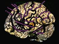 | The Vanderbilt Brain InstituteThe Vanderbilt Brain Institute is committed to activities that promote public awareness about the extraordinary advances in brain research and how those discoveries affect peoples lives. We coordinate Brainstorm a month-long series of brain awareness events for the public that focuses on how the brain works and the importance of brain research to understanding, treating, and ultimately curing brain related diseases. We invite brain science experts to Nashville throughout the year to share their discoveries. And we partner with non-profit associations and health advocacy groups to conduct brain education and outreach programs.
Click here for more information |  | Probability Density Volume for the CerebellumNovelty in the BrainMap database can be spotted using mathematical modeling of the activation foci. The novelty is detected as a discrepancy between the 3D coordinates and the anatomical label. The method finds a few entry errors in BrainMap together with a few errors in the original articles. The result is available as an automatic generated list of BrainMap outliers (Note the functional interface to the BrainMap database has been changed so the URL for the database does not work).Technical:The mathematical modeling is based on probability density modeling using the Specht kernel density estimate (Parzen window) applied on the 3D Talairach space conditioned on the words and phrases of the anatomical label. Novelty detection is implemented by directly ranking the likelihood.
Click here for more information |  | Awake brain mapping makes for some potentially entertaining surgeryBruce Hill was doing some Monday morning proselytizing about his Mormon faith, which is not unusual except that his skull was spread open and Dr. Mitchel S. Berger was tinkering with the temporal lobe.Hill was awake and talking during his brain surgery so that Berger could test his language skills. There was a tumor in there that had to come out. And the only way to know what brain matter could safely come out with it was to ask the patient. The procedure is called awake brain mapping and it works as a process of elimination. The surgeon uses an electrical charge to inactivate an area of the brain. Then the patient is presented with a question, in the form of a word or object drawn on a computer screen. If he can answer correctly with that area inactivated, then that tissue can be removed without causing brain damage.
Click here for more information | 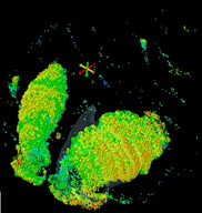 | Paul Allen's Digital BrainThis image shows expression of a specific gene, called etv1, in an area of the brain associated with motivation and reward. The color coding reflects the level of gene expression.
Click here for more information | 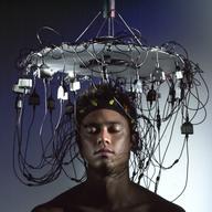 | EEG Performance Art
Click here for more information | 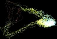 | EEG Attractor: Rat seizureAn EEG trace from the olfactory system of the brain of a rat, during an epileptic seizure. The trajectory of a point moving through this space in time traces the subspace that is occupied by a strange attractor. We have colored it red when the fourth variable is negative and blue-white when it is positive. The Hausdorff dimension is reduced to 2.52, and the structure of the strange attractor appears to be that of a 2-torus.
Click here for more information | 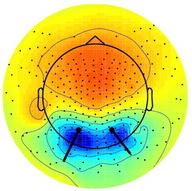 | Independent component of 252-channel EEG data with bilateral occipital scalp projectionDerived by independent component analysis of approximately 700,000 points of data using runica() in the EEGLAB Toolbox, implementing extended infomax ICA. Here, the 252-channel data were reduced to 160 independent components by PCA prior to ICA training. Channels located below the head center are shown in this topoplot() as extending out from the model head borders. Note that ICA finds the gradient of the projection from the source to the skin surface not only at the scalp electrodes but also for channels on the neck, forehead and temples. Residual variance of the bet-fiting symmetric dual-dipole model in a model sphere head was 2.0%. Dipole calculations used the DIPFIT plug in.
Click here for more information |  | The PsycographThe Psycograph was patented in 1905 by Henry Lavery of Superior WI. His first machine with 1,900 parts didn't work! A quarter century later, still building phrenology machines, Mr. Lavery recruited Mr. Frank P. White as an investor and began doing business as the Psycograph Company in the Builders Exchange, Minneapolis MN. Curator McCoy and woman in psychograph The psycographs were a novelty device featured in department stores and theatre lobbies during the Great Depression . The Psycograph Company operated from 1929-1937.
Click here for more information | 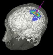 | Transcranial Magnetic StimulationTranscranial Magnetic Stimulation (TMS) consists of applying a focused magnetic field on a specific portion of the brain and of recording the reaction in another part of the body. In this example, the primary left motor cortex is stimulated and motor evoked potentials are recorded on the opposite hand.The figure shows interpolation of motor evoked potentials onto the brain surface. Center of gravity on the brain / scalp surface as well as at magnet level.
Click here for more information |  | Transcranial Magnetic StimulationFunctional brain mapping may be performed using a trans-cranial magnetic stimulation device. This coil generates magnetic field impulses which stimulate underlying nerve cells in a focused volume. By measuring responses to the stimulations and tracking the position of the coil relative to the MR scan we generate functional maps of the brain in a low-cost, non-invasive, and accurate manner. The LEDs placed on the face in the image below are used to track the position of the patient during the procedure, so that the patient's head does not have to be fixed.
Click here for more information | 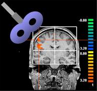 | Stimulating the Brain Makes the Fingers More SensitiveRepetitive transcranial magnetic stimulation (rTMS) has more than a whiff of Buck Rogers to it: a magnetic wand passes over the surface of the skull, triggering changes in the brain underneath. But it's not science fiction, and in the past decade, rTMS has emerged as an intriguing technique for exploring brain function, and a promising, though still unproven, form of therapy. Hubert Dinse, Martin Tegenthoff, and their colleagues show that a short course of rTMS can increase finger sensitivity for up to two hours after treatment ends, and that this change corresponds to an increase in the size of the brain map representing the finger.
Click here for more information | 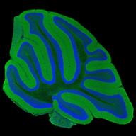 | Cerebellum Large Scale MosaicA high resolution multiphoton microscopy mosaic image of a cerebellar distribution of cell bodies (Hoescht 33342; blue) and alpha-synuclein (green) from a non-transgenic animal. These maps are being used to characterize Parkinson's Disease. These data will provide the bridge between whole brain imaging techniques such as MRI (Duke University, CIVM) and electron microscopic (UCSD-NCMIR) analysis.National Center for Microscopy and Imaging Research at UC San Diego
Diana Price, Ph.D
Mark H. Ellisman, Ph.D.
Maryann Martone, Ph.D.
Click here for more information | 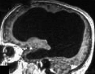 | Man with Tiny Brain Shocks DoctorsA man with an unusually tiny brain manages to live an entirely normal life despite his condition, which was caused by a fluid build-up in his skull.Scans of the 44-year-old man's brain showed that a huge fluid-filled chamber called a ventricle took up most of the room in his skull, leaving little more than a thin sheet of actual brain tissue. Intelligence tests showed the man had an IQ of 75, below the average score of 100 but not considered mentally retarded or disabled. "The whole brain was reduced frontal, parietal, temporal and occipital lobes on both left and right sides. These regions control motion, sensibility, language, vision, audition, and emotional and cognitive functions," Feuillet told New Scientist.
Click here for more information | 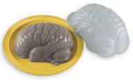 | Brain Gelatin MoldThink delicious!
Finally, what the world has been waiting for ... edible brains. This plastic mold produces the left hemisphere of a jiggly brain, about 7-1/2" x 6-1/2" -- larger than average for the general populace, but just right for Archie McPhee customers. Gelatin or aspic, your choice. Educational (you can identify the functions of the different parts of the brain as they slide down your throat), and fun (impress your friends as you re-enact scenes from "Night of the Living Dead"). Includes a recipe for brain-colored gelatin. After all, there's always room for brains.
Click here for more information | 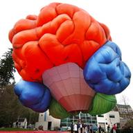 | Brain Awareness Week 2004BAW at the Oregon Health & Science University: Brain Balloon
Click here for more information |  | Brain of the Bottlenose DolphinThe Brain Museum web site provides browsers with images and information from one of the world's largest collection of well-preserved, sectioned and stained brains of mammals. Viewers can see and download photographs of brains of over 100 different species of mammals (including humans) representing over 20 Mammalian Orders.Also available are examples of stained sections from a wide variety of brains of special interest, including Humans, Chimpanzees, Monkeys, various Rodents and Carnivores, California Sealion, Florida Manatee, Big Brown Bat, American Badger, American Raccoon, Yellow Mongoose, Zebra, Cow, and the Atlantic Bottlenose Dolphin. A complete list of all available specimens is available. How brain evolution has occurred is discussed.
Click here for more information | 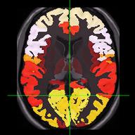 | PubBrain: Mental ImageryPubBrain is a project by Don Kalar that presents a graphical neuroanatomical access to the NLM's PubMed database. This image shows in color the frequency with which the given brain regions were named in papers whose abstract included the term, "mental imagery".
PubBrain is supported under the UCLA Center for Cognitive Phenomics (P20 RR020750).
Click here for more information | 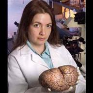 | The Dolphin BrainDr. Lori Marino with dolphin brain (Marino notes that all of her studies on dolphin brains are done on organs from animals that have died from non-research causes; e.g. strandings or disease). The brain of an adult bottlenose dolphin measures over 1,600 cc, versus the average adult human brain of about 1,450 cc. They have complex languages, social structures and, AM thinks, a voluminous memory for legends and sagas and knowledge passed down verbally from generation to generation. By killing dolphins, we destroy not only their biological bodies, but their cultures as well.
Click here for more information |  | Donner Brain MappingFrom the LBNL Picture archives. Donner brain mapping. In the Life Sciences Building, Campus, Dr. Edward Bennett (left), H. Morimoto and Marie Hebert measure enzyme activity in the brains of "bright" and "dull" rats.
Click here for more information | 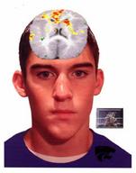 | K-State professor "maps" brains for effect of TV violence on youth"This isn't some isolated issue," said John Murray, a Kansas State University professor of family studies and human services. "This has been simmering for a long time and at some point we're going to have to come to grips with how we're going to deal with the ever-escalating violence."Murray and his colleagues anticipated and did see activation patterns that indicated emotional arousal while viewing the violence or activation of the limbonic system and general right hemisphere activations. What they did not expect to see was activation in two areas: the premotor cortex, an area on the right side of the brain that controls motor planning for action, and the posterior cingulate, which is reserved for long-term memory of traumatic events. According to Murray, the interpretation of the premotor cortex activation while viewing violence suggests that the subjects were attempting to imitate the boxing sequences.
Click here for more information | 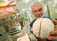 | Mapping Alcohol Brain DamageAssociate Professor Peter Dodd and his team from UQ's School of Molecular and Microbial Sciences has secured a five-year, $1.23 million grant from the United States' biomedical funding arm, the National Institute of Health (NIH) to find out.Chronic alcohol abuse kills brain cells in the front of the brain, which affect planning, and social interactions and inhibitions. The UQ team with collaborators from The University of Texas, want to find out what makes these cells so vulnerable. Professor Dodd said his team was analysing brain tissue from dead alcoholics, mostly Anglo-Celtic men who've averaged more than 10 drinks a day. The group analyses samples using a microarray analysis, a process they pioneered with human brain samples in 2000. From a DNA sample applied to a microscope slide, the microarray computer produces about 50,000 coloured spots showing which genes are expressed or switched on in the sample. What we're trying to find out is, what's different in the superior frontal cortex [front of brain] when you compare alcoholics with controls, that's not different in a spared area of the brain, Dr Dodd said.
Click here for more information |  | Manipulation of Brain Mapping ResultsBangalore, March 20The woman who conducted alleged rapist Abhishek Kasliwal's brain mapping test in Bangalore on Saturday claims three Congress men came to her soon after the test and asked her to manipulate the results.Dr S Malini, a forensic psychologist, said the men told her: "Please manipulate the results and certify Abhishek as innocent. His family's reputation and pride has taken a beating. The scientific tests have a lot of credibility and people will trust the report." Dr Malini, assistant director of the Forensic Science Laboratory, said she immediately ordered them to go away. Abhishek (26), the son of Shriram Mills owner Ambuj Kasliwal, has been accused of raping and assaulting a 52-year-old woman. "I told them to go away and not to return again with such requests. They said that Congress leaders from Maharashtra had contacted their leaders in Karnataka and sought help. I had to rudely show them the door," said Dr Malini. When asked if she found out more details about the men, she said, "I was very upset with them, so I did not continue the conversation." Reacting to the allegation, Karnataka Pradesh Congress Committee general secretary Prakash said, "We Congress-men never indulge in such acts. I do not know about the incident. If there is any specific complaint, we will look into it."
Click here for more information |  | Scientists are mapping the pathways that link emotion to health. The challenge for the rest of us is to put the discoveries to workNewsweek covers brain mapping
Click here for more information | 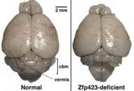 | Regulatory Pathway In Brain Development Possible Basis For MalformationsBruce Hamilton, Ph.D., associate professor in the Department Medicine, and his colleagues have identified a genetic regulatory pathway that controls a neural precursor cell's decision to self-renew as a dividing precursor or differentiate into a non-dividing neuron. Cells that are unable to differentiate appropriately and continue to proliferate may give rise to brain cancers. On the other hand, cells that differentiate too soon or make too few cells can result in malformations of critical brain structures.
Click here for more information | 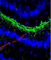 | Electrical Activity Alters Language Used By Nerve CellsUC San Diego biologists have shown that the chemical language with which neurons communicate depends on the pattern of electrical activity in the developing nervous system. The findings suggest that modification of nerve activity could have potential as a treatment for a wide range of brain disorders.In the study, published this week in the early on-line edition of the journal Proceedings of the National Academy of Sciences, the biologists showed that, contrary to the prevailing viewpoint, neurotransmitters--the chemical language of nerve cells--and receptors--the proteins that receive and respond to neurotransmitters--are not specified by a rigid genetic program. Altering nerve activity during development determines the "mother tongue" nerve cells use to communicate. The study will appear in the print edition of PNAS on January 2.
Click here for more information |  | Psychopharmaceuticals and brain imaging could make prisoner interrogation more humane. Should we use them?From Slate.comAbu Ghraib represents far more than disgraceful lapses in military supervision and honor. Its all-too-graphic institutional excesses signal that America must consider different ways to procure valuable information from terrorists, unlawful combatants, and insurgents.The good news is those ways exist, or will soon. Interrogators will soon be able to secure reasonably reliable information from their detainees without harsh sleep deprivation, physical threats, or sexual humiliations. The rationaleand the rationalizationsfor outrageous abuse will dissolve.
Click here for more information |  | BSM Mind Alertness ProgramA clever ad campaign for a program specializing in driver's education
Click here for more information | 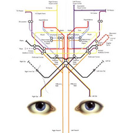 | Mapping the Mind, by Rita CarterIn the last decades of the 20th century, scientists have come to believe that the human brain is almost completely modular. Every bit of the brain does something in particular, and surprisingly specific abilities, memories, and responses are in localized areas. Journalist Rita Carter has drawn a map of what is known (and speculated) about the mind in a heavily illustrated field guide to the human brain.Carter and her scientific editor, neuropsychologist Christopher Frith, cover the state of the mind in a reasonably accurate, accessible way. They emphasize topics that are likely to be of some practical interest--such as Alzheimer's or attention deficit disorder--but not so much as to give a distorted picture of the field. Perhaps the most interesting parts of the book are the sidebars written by a variety of leading names in mind-brain science. Roger Penrose writes on computer minds, Francis Crick on consciousness, Steven Rose on memory, John Maynard Smith on social evolution, William Calvin on mosaic minds, Kay Redfield Jamison on creativity and bipolar disorders, and more. It's a stellar assortment, more than worth the price of admission--and there's a map of the mind on the cover, in case you misplace yours. --Mary Ellen Curtin
Click here for more information |  | Cat brain mapCredited to: Earl Blockenfeld http://radio.weblogs.com/0107064/
Click here for more information |  | Robotic arms for brain surgeryA pair of robotic arms capable of performing delicate operations on
the brain. Developed by a team at the University of Calgary, Canada,
the neuroArm allows surgeons to perform remote procedures on patients
lying in MRI scanners, to get the clearest 3D image possible of even
the tiniest nerves.
Click here for more information |  | Drosophila CortexA much-needed resource for the continued understanding of Drosophila memory formation is a high-resolution map of the brain. Ron Davis' laboratory are using two-photon microscopy to visualize neurons in the Drosophila brain and are developing the computational and software tools necessary for constructing a digital map of the brain and for analyzing brain organization within and between individual animals.
Click here for more information | 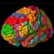 | Brodmann Areas on Colin BrainRendered by Mark Dow
Click here for more information |  | Association fibre pathways of the brainAssociation fibre pathways of the brain: parallel observations from diffusion spectrum imaging and autoradiography
Understanding the long association pathways that convey cortical connections is a critical step in exploring the anatomic substrates of cognition in health and disease. Diffusion tensor imaging (DTI) is able to demonstrate fibre tracts non-invasively, but present approaches have been hampered by the inability to visualize fibres that have intersecting trajectories (crossing fibres), and by the lack of a detailed map of the origins, course and terminations of the white matter pathways. We therefore used diffusion spectrum imaging (DSI) that has the ability to resolve crossing fibres at the scale of single MRI voxels, and identified the long association tracts in the monkey brain. We then compared the results with available expositions of white matter pathways in the monkey using autoradiographic histological tract tracing. We identified 10 long association fibre bundles with DSI that match the observations in the isotope material: emanating from the parietal lobe, the superior longitudinal fasciculus subcomponents I, II and III; from the occipital-parietal region, the fronto-occipital fasciculus; from the temporal lobe, the middle longitudinal fasciculus and from rostral to caudal, the uncinate fasciculus, extreme capsule and arcuate fasciculus; from the occipital-temporal region, the inferior longitudinal fasciculus; and from the cingulate gyrus, the cingulum bundle. We suggest new interpretations of the putative functions of these fibre bundles based on the cortical areas that they link. These findings using DSI and validated with reference to autoradiographic tract tracing in the monkey represent a considerable advance in the understanding of the fibre pathways in the cerebral white matter. By replicating the major features of these tracts identified by histological techniques in monkey, we show that DSI has the potential to cast new light on the organization of the human brain in the normal state and in clinical disorders.
Click here for more information |  | PhotoFrom the Flickr user known as Brain MapShe writes, "The mind is its own place, and in itself
Can make a heaven of hell, a hell of heaven." I am five feet, five inches tall.
During my adolescence, I lived in a home with fish,snakes,iguanas, rats, hamsters, cats, and dogs; we should've charged admission.
On weekends, I refuse to wake up until the hour number reaches the double digits.
I don't know how to ride a bike.
I think of the philosophy within the "Tao Te Ching" as the best advice I've ever heard, and I wish I was better at following it.
It makes me happy when I look out the window and see rain.
I've lived in California all my life but want to live in wildly different environments in the future.
My father was raised in Peru, my mother in a small mountain town near San Diego.
My favorite genre of food is Indian but if I had to eat one thing for the rest of my life it'd probably be nachos.
I have an inexplicable love for red shoes, especially when they provide the only red within an outfit.
I used to often dream about losing my teeth but now rarely dream at all.
I don't like strawberries but I love strawberry-flavored foods.
My boyfriend has a big, soft heart.
I'm Female and Taken.
Click here for more information |  | Sensory Map of C. ElegansElizabeth Ryder's group are studying the formation of a very simple sensory map in C. elegans. The six neurons in the map, known as the IL2 neurons, each send sensory processes to the worm's nose and axons to the nerve ring (the worm's "brain"). The entire map can be visualized in living animals using the fluorescent dye, DiO. By screening for mutations that disrupt this map, we have isolated several alleles of a gene called dig-1. Sensory processes of dig-1 mutants often follow aberrant paths to the worm's nose, sometimes branching abnormally . The dig-1 gene appears to be a very large molecule that is involved in adhesion. They are currently characterizing the expression pattern of this gene using molecular techniques. Mutations in the gene mig-10 also disrupt the IL2 neurons. In this case, the axons rather than the sensory processes are affected, sometimes stopping before reaching the nerve ring. We are also analyzing the expression pattern of the mig-10 gene. Future work will involve additional genetic screens to isolate and characterize mutations in other genes involved in sensory map formation in the C. elegans nervous system.
Click here for more information |  | Zernov's EncephalometerThe first prototype of a stereotaxic instrument was conceived and designed in 1889 by Dmitrii Zernov (1843-1917) - a professor of anatomy at Moscow University. His invention was based upon the similarity of the shape of cranium and brain to the terrestrial globe. This resulted in a real geographical atlas of the human brain, where each cerebral structure was expressed in the degrees of longitude and latitude. Zernov wrote: "I built an instrument which enables to project the pattern of cranial sutures or cerebral sulci or deep-seated brain structures on the spherical surface and then transform it onto the plane similar to the projection of the terrestrial globe on the map. The localisation of a given point on the brain surface is determined by the degrees of latitude and longitude."
Click here for more information | 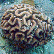 | Brain CoralThe beaches on Maui were gorgeous. On the first day of our vacation I happened to spy what appeared to be a bit of brain coral on the ocean floor a ways out from shore; this intrigued me. Wild coral! We bought goggles, and on the second day I swam out to get a better look.
The waves that day were rather large, but not so large as to cause concern. I was swimming around, minding my business looking for the coral when an errant wave picked me up. Well, I found what I was looking for! The coral opened up my leg (warning: blood) a little bit. It also left a little bit of itself in me. This was about two weeks ago.For a few days I tried to dig the shard out of my left big toe, but I just couldn't seem to pull it out. I eventually gave up and let it go, hoping it would work itself out. As it turns out, it's really annoying to have a piece of something embedded in one's toe! Today I went to the hospital to see what could be done about the fact that I was carrying a bit of Hawaii with me everywhere I went. Me: "I have a rock in my toe." Doctor: "Ah, I see infection. Antibiotics, and injection. Tetanus injection." Me: "Yes, it's a little infected. I had a tetanus shot last year. The rock is still in my toe." Doctor: "So antibiotics five days."
Me: "The rock is in my toe." Doctor: "Rock? Now?" Me: "Yes." Doctor: *sends me to get X-rays* Me: *gets X-rays* Doctor: "There is rock in your toe." The X-rays showed that a shard of coral had entered my toe and struck the bone, fragmenting. I had minor surgery to have the shards (one 1-cm bit, and three tiny fragments) removed; there was some between-the-toe syringe action, then some scalpelin', and some suture-slingin'. (Total cost for consulting, two X-rays, OR fees and surgery with anesthetic and sutures, plus antibiotic injection: about $40.) Now, thankfully, my days as an endogenous coral smuggler are over and I have the fragments as external souvenirs. Hooray!
Click here for more information |  | Jeff Hawkins - Founder of NumentaJeff Hawkins (born June 1, 1957 in Huntington, New York) is the founder of Palm Computing (where he invented the Palm Pilot) and Handspring (where he invented the Treo). He has since turned to work on neuroscience full-time and has founded the Redwood Neuroscience Institute and published On Intelligence describing his memory-prediction framework theory of the brain. In 2003 he was elected as a member of the National Academy of Engineering "for the creation of the hand-held computing paradigm and the creation of the first commercially successful example of a hand-held computing device."
Click here for more information | 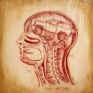 | I drive, therefore I am.Artwork by Alie Ward
Click here for more information | 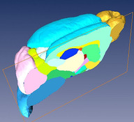 | Sagittal cut through the 3D microMRI C57BL/6J mouse brain atlasThe image shows in great detail a sagittal cut through the 3D microMRI C57BL/6J mouse brain atlas. Brain structures are labeled in different colors. The structures visible here are neocortex (cyan), olfactory bulb (golden), cerebellum (pink), brain stem (blue), caudate putamen (light green), hippocampus (deep blue), thalamus (milky white), external capsule (yellow), internal capsule (red), globus pallidus (deep pink), amygdala (light yellow), inferior coliculli (yellowish green), superior coliculli (white) and hypothalamus (deep cyan). Image courtesy of Dr. Helene Benveniste and Dr. Yu Ma, Brookhaven National Laboratory and Stony Brook University. Grant NIH R01 EB 00233-04 and P41 RR16105.
Click here for more information | 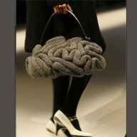 | Brain BagThe perfect accessory for the serious scientist.
No Brainer
Is it a hovercraft?
Is a monster?
Is it alive?
Presenting the Undercover Brain Bag!
Feel it, hold it and fall in love with the brain- only one surreal lonesome available at Someday!
Click here for more information |  | MRC Cognition and Brain Sciences UnitThe unit's research programme investigates fundamental human mental processes such as attention, memory communication and emotion. We conduct behavioural experiments to determine how these processes work, build computer models of their operation, study how they could arise from the neural mechanisms of the brain, and explore the clinical implications of al our studies for patient therapy and rehabilitation.
Click here for more information | 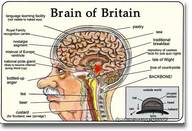 | Brain of BritainNeuroimaging Britishness:
A recent study comparing British and non-British participants has found some compelling differences in brain structure that may account for differences in national character.
Click here for more information | 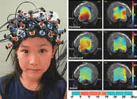 | Commercial Mind-Machine Interface by 2011Hitachi's new neuroimaging technique allows its operator to switch a train set on and off by thought alone, and the Japanese company aims to commercialize it within five years.
Click here for more information |  | Yale Developmental Neuroimaging ProgramThe Developmental Neuroimaging Program (DNP) is a newly launched initiative designed to foster collaborative science on the development of the brain, and brain-based neuropsychiatric disorders. The program is co-directed by Robert T. Schultz, PhD, of the Child Study Center and Lawrence Staib, PhD, of Diagnostic Radiology, and it includes multiple adjunct faculty throughout the University. The Program cuts across existing laboratories within the University and brings together faculty with complementary areas of expertise in order to provide an arena for collaborative research and training in neuroscience.
Click here for more information |  | Faith and Healing by John HerseyFrom an illustration in The New York Times
Click here for more information |  | Whole Brain Model - HerrmanWhat is the whole brain model?The Whole Brain Model from Ned Herrman is a technique that can be used for analyzing personal and organizational thinking preferences.The Whole Brain Model is a mental model that describes thinking preferences. These are the ways of thinking that satisfy us the most and seem natural for us at this point in our lives. These ways of thinking can change, often as a result of significant emotional experiences, life transitions and other important insights. Thinking preferences describe the patterns of what we prefer to pay attention to and what we don't prefer to pay attention to.
Click here for more information |  | Your Brain is Not Your FriendA mind is a terrible thing. Whether because of the brains internal structure or the way social and cultural pressures cause our minds to develop and function, in the end the result is the same: minds that are not only easily deceived and frequently deceptive in their own right, but when caught out, refuse to accept and address their errors. If you have a mind or even half a mind you might be best off losing it entirely. Barring that, though, there are a few things you should know about the enemy in your head. Before it hurts someone
Click here for more information |  | Protecting the Brain with ProgesteroneIn a small, preliminary study of 100 patients published recently in the Annals of Emergency Medicine, doctors gave brain injury patients high intravenous doses of the pregnancy hormone progesterone within 12 hours of injury. Patients who received the hormone cut their risk of dying by 57 percent. The study "gives hope where there has been no hope before," said the the study's lead author, Dr. David Wright, an assistant professor in the department of emergency medicine at Emory.
Click here for more information | 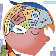 | The New Yorker maps the BrainA particularly appealing New Yorker cover from the September 4, 2006 issue.The original image can be found [here].
Click here for more information |  | CTF MEG Magnetoencephalography Brain Imaging SystemMagnetoencephalography (MEG) is a revolutionary medical imaging technology that provides unprecedented insight into the workings of the human brain through the measurement of electromagnetic activity.By measuring the magnetic fields created by the electric current flowing within the neurons, MEG identifies brain activity associated with various human functions in real time, with millimeter spatial accuracy. This non-invasive approach can positively impact patient outcomes, providing clinicians with the invaluable information they need to evaluate neurological disorders and plan surgical treatments. Our CTF MEG systems are the most technologically advanced instruments available in the world today, offering MEG sensor arrays of up to 275 distinct channels with up to 128 simultaneous EEG sensors. The exquisite sensitivity of the MEG sensors is achieved by utilizing the world's most sensitive detectors of magnetic fields, SQUIDS (Superconducting Quantum Interference Devices)
Click here for more information | 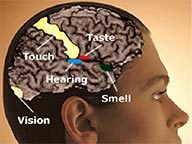 | Hearing Beautiful Paintings - A Relectionist Informs NeuroestheticsAs neuroesthetics, the study of the neurobiology of artistic creativity and achievement, continues to expand, new forms of art like Marcia's will emerge at the nexus of our new knowledge of brain and what it can create. Just as information technology has made new forms of art possible like brain wave synthesizers, digital banners and electronica, neurotechnology will surely play an important role in the ever evolving world of art, architecture and entertainment in the years to come.
Click here for more information | 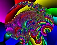 | And this is your brain on drugsThe fractals here were created by Sven Geier, who is currently employed by the Jet Propulsion Laboratory (JPL); a division of the California Institute of Technology and a center of the National Aeronautics and Space Administration (NASA). He lives in Pasadena, CA, with his wife, his little son and two cats. In his copious spare time he produces amongst other things web-pages like this one and artwork like what you see here. "I created these because it pleases me to create them. Go ahead and download them, save them, give them to your friends, print them and hang them on a wall, whatever you want. "Property" is simply not a productive paradigm for art." If you like these images and you do something special with them, I'd be glad if you could drop me an e-mail, though, as I like getting feedback. And of course you're allowed to donate, if you'd like.
Click here for more information | 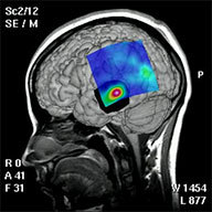 | Nonlinear EEG Analysis of Epilepsy PatientsEpilepsy manifests itself not only in visible seizures, but also in characteristic patterns in the electroencephalogram (EEG). It is thus an important field of application for modern methods of nonlinear time series analysis. In collaboration with the Department of Epileptology at the University of Bonn, we are pursuing two main aims. On the one hand, we develop methods for localizing epileptic foci in patients who are possible candidates for surgery. The figure shows regions with elevated (green) and high (red) likelihood of being a focus in a retrospective analysis. Also shown is the region which has actually been removed, based on other analyses (black). In the case illustrated, the primary focus had been correctly recognized and removed, and the patient is now free of seizures. An advantage of our method over previous ones is that it uses only data from seizure-free epochs, and that their predictions do not always agree with those of other methods. In some rare cases where the patient is not completely free of seizures, our analysis would suggest that there is indeed a secondary focus or that the primary focus has not been removed entirely.
Click here for more information |  | Positioning of EEG electrodes on the cerebral cortexAn EEG is a record of brain activity that is measured using an electroencephalograph and a series of electrodes. In order to perform consistent testing of electrical brain activity, the International Electrode Placement System or 1020 system was developed in 1947. The system is based on the relationship of the location of the electrode and the underlying area of cerebral cortex. It takes into account varying head sizes by measuring the lateral distance between the ears and the longitudinal distance from the bridge of the nose to the back of the head, with electrodes being placed at 10 and 20 per cent values of a measured distance. Each site on the scalp has a letter (to identify the lobe) and a number or another letter to identify the hemisphere location. Electrodes positioned over the frontal (F), temporal (T), parietal (P) and occipital (O) lobes are represented by the corresponding letter. Odd (1, 3, 5, 7) and even (2, 4, 6, 8) numbers refer to the left and right hemispheres, respectively. The closer the position is to the midline, the smaller the number. A 'z' refers to an electrode placed on the midline.High resolution version
Click here for more information | 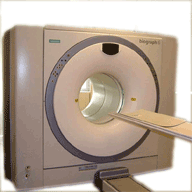 | PET/CTPET/CT is a new imaging tool that combines two scan techniques in one exam - a PET scan and a CT scan. PET/CT is mainly used for diagnosis, staging or restaging malignant disease and metastases and evaluation of treatment response. It may also be used to differentiate dementia verses Alzheimer's disease. The two procedures together provide information about the location, nature of and the extent of the lesion. In other words, it answers questions like: Where is the tumor, how big is it, is it malignant, benign or due to inflammatory change, and has the cancer spread?
Click here for more information |  | Neuroimaging Tools Available On WebA roving band of five unnamed researcher participants-who traveled across the country to nine different sites to have their brains examined via MRI-has contributed to a first-of-its-kind neuroimaging dataset that will help researchers to standardize and calibrate imaging data for multisite studies for years to come. The dataset, known as the Function BIRN Phase I Traveling Subjects Dataset, is the latest of more than two dozen open-source data and software tools made available to researchers worldwide by the Biomedical Informatics Research Network (BIRN).
Click here for more information | 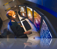 | Neuroimaging at Texas A&MThe head of the College of Medicine's department of psychiatry, Kathryn Kotrla, M.D., is pursuing new techniques in neuroimaging--the use of magnetic resonance imaging (MRI) machines to view what is happening in the brains of people with severe mental illnesses such as schizophrenia. MRI can now be used to create "movies" of processes within parts of the body, including the brain. Some of the newer applications of magnetic resonance imaging include functional MRI (FMRI)--in which brain activity can be viewed as it happens--and magnetic resonance spectroscopy (MRS), which allows researchers to discern the concentrations of various chemicals in the brain.
Click here for more information | 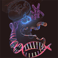 | Brainethics - Consequences of Brain ScienceScience and technology always work in tandem. Neurotechnology refers to the set of tools that have been developed to analyze and influence the human nervous system, especially the brain. We would like to assess the uses that are or could in the future be - made of new neurological knowledge and technologies. What are the consequences when biochemical solutions to behavioral problems such as depression, addiction, or eating disorders take precedence over attempts to repair the social environment, or defective inter-personal relations? How do we avert the risk of psychopharmacology being abused for neurochemical enhancement?
Click here for more information | 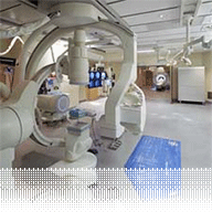 | TWH opens Canada's top neuroimaging deptTWH Neuroimaging Department features the worlds only integrated 3T MRI unit, biplane angiographic suite and OR Combo Room, Ontarios only Gamma Knife and Canadas first 64-slice CT scanner. Shown here: Angiography suite, complete with integrated OR and 3T MRI.
Click here for more information | 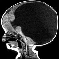 | Holoprosencephaly NeuroimagingNEUROIMAGING EVALUATION of HPE is best accomplished by MRI. In all suspected cases the scan should include axial, sagittal and coronal sequences to enable complete evaluation of midline structures. Also, thin sections are recommended; ideally no greater than 3 mm thick, with spacing of 0.5 mm. Even better are volumetric protocols, which generally have 1.5 mm slice thickness with no gaps.Until now, a lack of standardization of protocols has made it difficult to meaningfully classify this disorder. The individuals to the right have been instrumental in helping the Carter Centers to develop standardized MRI sequences to obtain 3-dimensional structural data for HPE, which can then be compared across many institutions. It is hoped that utilization of improving technology to fully characterize brain structure in HPE will lead to better classification schemes, important genotype-phenotype correlations, and ultimately to a better understanding of etiologies.
Click here for more information |  | A brain-computer interface (BCI)This brain-computer interface (BCI) system assists patients who are completely paralyzed from severe neuromuscular disorders such as amyotrophic lateral sclerosis (Lou Gehrig's disease), brainstem stroke, or high-level spinal cord injury. In a BCI system, the brain's electrical activity is detected through the scalp and processed by a computer to extract wave patterns indicating the user's intent. The patterns are then translated into commands for a device, such as moving a cursor on a computer screen or operating a wheelchair. Image courtesy of Dr. Jonathan Wolpaw, Wadsworth Center, Albany, New York. Grant EB000856
Click here for more information | 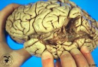 | Brain Damage and Language Comprehension: Wernicke's AphasiaVideos of aphasia
Click here for more information | 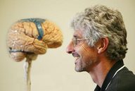 | Award opens up learning opportunities for medical imaging expertDoctoral student Michael Chappell is looking forward to brainstorming with international researchers in Italy this June at an international neuroimaging conference.
Click here for more information | 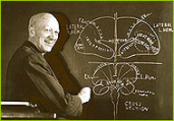 | Wilder PenfieldWilder Penfield, a pioneering brain surgeon, mapped the motor cortex using mild electric current. This is a link to the Nova "Secrets of the Mind" web site
Click here for more information | 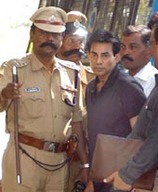 | Salem brought to Bangalore for testsBangalore, December 28
Extradited underworld don Abu Salem was brought here today under tight security to undergo brain-mapping, narco-analysis and polygraph tests at the forensic science laboratory. Flown in by Mumbais anti-terrorist squad (ATS), Salem was brought here by a team led by Deputy Inspector-General of Police Jai Jeet Singh to ensure he is not targeted by rival gangs.
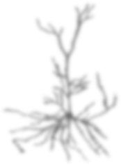
Click here for more information |  | Quantum Power Chip Immediately Increased Energy to all Parts of the BrainClick here for more information |  | Sculpting the brainscapesAn ink drawing form \"Glia and Muscle Sculpt Neuromuscular Arbors by Engulfing Destabilized Synaptic Boutons and Shed Presynaptic Debris\", which appeared in PLoS Biology (see Fuentes-Medel et al., e1000184) showing that synaptic growth at the neuromuscular junction entails substantial shedding of membranes, which are removed through a collaboration between muscles and glial cells.
Image Credit: Vivian Budnik, University of Massachusetts Medical School
Click here for more information | 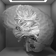 | Transparent FibersYoshihito Yagi is exploring sophisticated rednering methods for displaying the results of diffusion tractography. HIs 2005 master's project, Visualizing Fiber Tracts in the Brain
Using Diffusion Tensor Data was the development of FiberKit, a toolset for interactive visualization of these brain connectivity data.
Click here for more information | 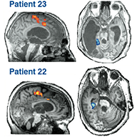 | Sculpting the brainscapesAn ink drawing form \"Glia and Muscle Sculpt Neuromuscular Arbors by Engulfing Destabilized Synaptic Boutons and Shed Presynaptic Debris\", which appeared in PLoS Biology (see Fuentes-Medel et al., e1000184) showing that synaptic growth at the neuromuscular junction entails substantial shedding of membranes, which are removed through a collaboration between muscles and glial cells.
Image Credit: Vivian Budnik, University of Massachusetts Medical School
Click here for more information |  | Virgin Mary in Fort Pierce woman's brain scanFORT PIERCE A 42-year-old woman without insurance and mounting medical bills plans to sell an MRI scan of her brain in which the image of the Virgin Mary seems to appear.
Pamela Latrimore has been sick for years with cancer, arthritis and a series of serious ailments she blames on a childhood in Jacksonville, Ark., a place that has been investigated by the U.S. government for possible dioxin exposure. Dioxin is a toxic chemical linked to a variety illnesses including cancer and liver problems.
In 2002, Latrimore had an MRI of her brain done and the results were stashed in her thick pile of medical records. Her sister-in-law looked at the sheet recently and pointed out what appeared to be the image of the Virgin Mary.
Having seen where other supposed images of Mary or other religious icons were sold for thousands of dollars, Latrimore plans to post the MRI scan on eBay, the online auction site. She hopes to earn enough money to pay off some of the medical bills she and her contractor husband cannot afford.
Click here for more information | 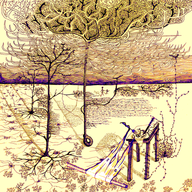 | Sculpting the brainscapesAn ink drawing form "Glia and Muscle Sculpt Neuromuscular Arbors by Engulfing Destabilized Synaptic Boutons and Shed Presynaptic Debris", which appeared in PLoS Biology (see Fuentes-Medel et al., e1000184) showing that synaptic growth at the neuromuscular junction entails substantial shedding of membranes, which are removed through a collaboration between muscles and glial cells.
Image Credit: Vivian Budnik, University of Massachusetts Medical School
Click here for more information |  | Researchers used fMRI technology to try to pull images out of peoples' brains. Having modeled how images are represented in the brain, the researchers translated recorded patterns of neural activity into pictures of what test subjects had seen. Though practical applications are decades away, the research could someday lead to dream-readers and thought-controlled computers. "It's what you would actually use if you were going to build a functional brain-reading device," said Jack Gallant, a University of California, Berkeley neuroscientist. The research, led by Gallant and Berkeley postdoctoral researcher Thomas Naselaris, builds on earlier work in which they used neural patterns to identify pictures from within a limited set of options. The current approach, described this week in Neuron, uses a more complete view of the brain's visual centers. Its results are closer to reconstruction than identification, which Gallant likened to "the magician's card trick where you pick a card from a deck, and he guesses which card you picked. The magician knows all the cards you could have seen." In the latest study, "the card could be a photograph of anything in the universe. The magician has to figure it out without ever seeing it," said Gallant.
Click here for more information |  | H.M. dies at 82Henry Gustav Molaison, known to the science community as H.M. passed away today (December 4th, 2008) due to respiratory failure. H.M. will forever be remembered for his immeasurable contribution to our understanding of the brain and memory. On september 5, 1953 H.M. underwent medial temporal lobe surgery for the treatment of intractable epilepsy. In the following months H.M.'s seizures resolved, but curiously he was left with anterograde amnesia; leaving unable to form new long-term memories. In the following years, up until this past week scientist have continuously studied H.M. attempting to better understand how memories are formed and retrieved. Specifically, work with H.M. has helped us understand the brain's declarative versus working memory systems. Even more importantly, H.M. will be remembered for his kindness and wiliness to act as a research subject for the advancement of science.
Click here for more information |  | Sculpting the brainscapesAn ink drawing form \"Glia and Muscle Sculpt Neuromuscular Arbors by Engulfing Destabilized Synaptic Boutons and Shed Presynaptic Debris\", which appeared in PLoS Biology (see Fuentes-Medel et al., e1000184) showing that synaptic growth at the neuromuscular junction entails substantial shedding of membranes, which are removed through a collaboration between muscles and glial cells.
Image Credit: Vivian Budnik, University of Massachusetts Medical School
Click here for more information | ![]() | Sculpting the brainscapesAn ink drawing form \"Glia and Muscle Sculpt Neuromuscular Arbors by Engulfing Destabilized Synaptic Boutons and Shed Presynaptic Debris\", which appeared in PLoS Biology (see Fuentes-Medel et al., e1000184) showing that synaptic growth at the neuromuscular junction entails substantial shedding of membranes, which are removed through a collaboration between muscles and glial cells.
Image Credit: Vivian Budnik, University of Massachusetts Medical School
Click here for more information | ![]() | qqqzsdz
Click here for more information |  | qqqzsdz
Click here for more information | ![]() | qqqsdssddsssssssss
Click here for more information |
|

