Interval discrimination across temporal scales relies on different neural circuits: an event-related fMRI study
J.E. Bramen; D.V. Buonomano; M.S. Cohen. Brain Mapping Center, UCLA School of Medicine, Los Angeles, CA 90095
to be presented at the Society for Neuroscience, 2005
Sunday, Oct. 24, 9:00 AM - 9:15 AM
Session Number: 138.5 Location: Room 7B
Temporal processing occurs over a large range of time scales, including perceptual timing (tens to hundreds of ms) and time estimation (seconds). It is not known whether the same or different brain regions are responsible for these different forms of temporal processing. The purpose of this study is to assess whether differing time scales rely on the same neural circuitry. Because dissociations exist in the type of timing affected by different lesions, we expected differences in brain activation patterns across scale. These data support this hypothesis
Six subjects performed auditory temporal and frequency discrimination tasks during event-related fMRI. Subjects were presented with three blocks of 40-paired tones. In the interval conditions, subjects judged whether comparison intervals were longer or shorter than 100 ms (short interval) or 1200 ms (long interval). In the frequency condition subjects decided whether the pitch of comparison tones were higher or lower than a 3 kHz standard.
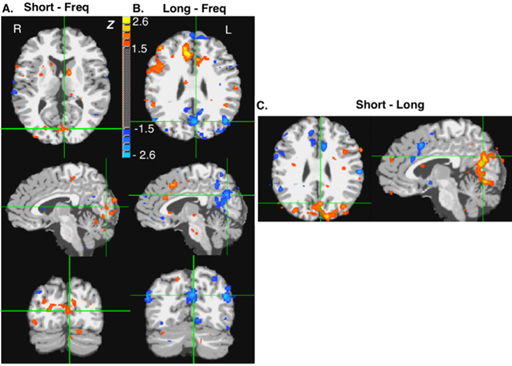
Both the short and long interval conditions were assessed relative to frequency. Differences between the two conditions include the following: The anterior cingulate gyrus, anterior lobe of the cerebellum, SMA, L pulvinar and R substantia nigra were active in the short but not the long interval condition. The parietal-occipital sulcus and calcarine fissure were activated in the short but deactivated in the long interval condition. The R IFG and R MFG were active in the long but not the short interval condition. Overlap between short and long interval conditions relative to frequency exists in the caudate, globus pallidus, insula, pontine nuclei, and R SPL.
These data support the hypothesis that temporal processing on different scales does not rely on common centralized brain regions.

Comparison of visual regions in 100 ms and 1200 ms interval conditions. All contrasts thresholded for z values of magnitude greater than 1.5. A) Regions whose MR signal differed in the 100 ms interval discrimination compared to the frequency control include cuneus, precuneus, inferior middle and superior occipital gyri. B) Regions whose MR signal was reduced in the 1200 ms interval discrimination compared to frequency included the cuneus, precuneus, middle and superior occipital gyri. C) Regions whose MR signal differed in the 100 ms interval condition compared to the 1200 ms interval discrimination.
|

