
| Faculty | Contact |
FAQ | Home | Summer Program |
neuroscience, magnetic resonance, functional mri, fMRI, neurosurgery, brain mapping, cognitive neuroscience, eeg, positron emission tomography, ucla, neuroimaging, pulse sequences, alzheimer, parkinson, perception, neurophysiology, magnetic resonance imaging, functional magnetic resonance imaging, cognition, psychology, epilepsy, PET, optical intrinsic signal, OIS, QEEG, magnetoencephalography, MEG, spectroscopy, nih, nimh, spm, fsl, afni, macine learning, physics. electroencephalography, electrophysiology, imaging, rf coil, diffusion, tractography, connectome, big data
NITP Fellowship Awardees
updated 1/28/11
Publications by the NITP Awardees
 |
Nanthia Suthana, Ph.D.My primary research focus is on the neural basis of learning and memory through combination of single-unit recordings with high-resolution magnetic resonance imaging (MRI) in order to record from neurons within hippocampal subregions during learning and memory. I am also investigating modulation of memory in temporal-lobe epilepsy patients in which deep brain stimulation is currently underway. The NITP Provided a multimodal foundation of Neuroimaging as a tool for studying cognitive function. Resources were extremely useful in designing, implementing, analyzing, and interpreting research projects. Faculty were extremely open, helpful, and collaborative. Lastly, courses were fun as well! |
|
Present Position: Postdoctoral fellow. PI: Itzhak Fried / Barbara Knowlton. Cognitive Neurophysiology Laboratory. UCLA Department of Neurosurgery
|
||
Elliot Berkman, Ph.D.How do we pursue long-term goals? What are the behavioral, motivation, and neural systems that contribute to our success or failure? A central aim of Dr. Berkman’s research is to understand how these systems work together to help us pursue our goals. To do this, he combines the distinct strengths of several research methods including functional magnetic resonance imaging (fMRI), cross-sectional and longitudinal survey methods, and laboratory experiments. Examples of his research include fMRI studies of basic goal-relevant processes such as self-regulation and inhibitory control, experimental studies on how approach and avoidance motivation relate to emotions and performance, and longitudinal studies on real-world goals such as smoking cessation. As part of this research program, Dr. Berkman is developing and using new statistical techniques to integrate data across a number of methodologies, including structural equation modeling, idiographic analysis of fMRI data, and joint hierarchical linear modeling of fMRI and behavioral data. |
 |
|
The neuroimaging training that I received during NITP was a considerable factor in my offer for a tenure-track assistant professor from University of Oregon. They were looking for someone who could teach statistics and neuroimaging; thanks to NITP I became far more expert in both than I otherwise would have been. They were especially impressed by my facility developing custom code for fmri analysis--something that I began to learn because of NITP and have continued to develop for the duration of my graduate career. Present Position: Assistant Professor. Director, Social Affective Neuroscience Laboratory. University of Oregon
|
||
 |
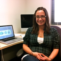
Pamela Douglas, Ph.D.Dr. Douglas is a biomedical engineer with a strong interest in quantitative analysis of neurobiological and neuroimaging data and particular attention to machine learning. Her work since the NITP includes three full journal publications and five meeting abstracts. |
|
Present Position: Graduate student researcher, laboratory of Susan Bookheimer, UCLA
|
||
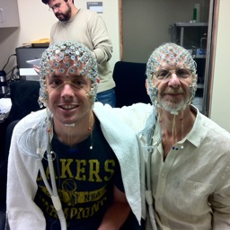
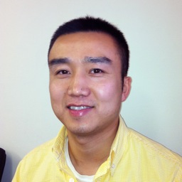
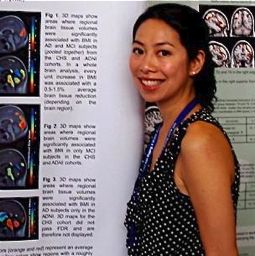
Jesse Brown (left)My research is in multimodal neuroimaging of human episodic memory systems in health and neurodegeneration. Currently, I use DTI and fMRI to study connections between the hippocampus and the prefrontal cortex in normal individuals and those at greater genetic risk for developing Alzheimer's Disease. More generally, I'm interested in memory, early detection of neurological disease states, human brain evolution, and cognitive enhancement/augmentation. The NITP prepared me to perform multimodal analysis with fMRI, DTI, and EEG scans. The benefits of this extended program are now coming around. I was second author on a DSI/DTI reproducibility paper in Neuroimage, for which I wrote the software to analyze this data. The programming experience I gained in the NITP was integral to this work. The NITP also provided me with the knowledge to TA for the UCLA Advanced fMRI summer course, presenting lab sections on both DTI and fMRI connectivity analyses. I have also met numerous well-respected and well-known professors in the last two years through their affiliation with the NITP summer program, forming critical connections and receiving multiple postdoc offers. |
 |
|
Present Position: Postdoctoral fellow, laboratory of Mark Cohen, UCLA.
|
||
 |
Doris Payer, Ph.D.I am interested in understanding the neuro-cognitive correlates of stimulant abuse, and their relationship to impulsive and compulsive behaviors that can perpetuate addiction. |
|
Present Position: Post-doctoral fellow, Centre for Addiction and Mental Health, Addiction Imaging Research Group, Toronto, ON, Canada
|
||
Jason SteinThe structure of the brain is largely based on genetic factors, but the specific variants which account for this high heritability are mostly unknown. We are seeking common genetic variants which account for some portion of the heritability of brain structure using genome-wide association. We have explored associations using multiple phenotypes including temporal lobe volume, caudate volume, and hippocampal volume. To have sufficient power to reliably identify variants of small effect, we recently formed a large multi-national organization called the Enhancing NeuroImaging Genetics through Meta-Analysis (ENIGMA) consortium to create a meta-analytic imaging genomics network. The first project of the ENIGMA consortium is currently underway and is conducting a GWAS meta-analysis using hippocampal volume as a phenotype with a projected N approximately 7000. Variants identified using brain structure as a phenotype may provide an alternative to using heterogeneous clinical disease categories for gene-hunting expeditions. |
 |
There is no other class like the NeuroImaging Training Program to prepare oneself for research in the neuroimaging field. The wide array of subjects covered helped put into perspective the neuroimaging field as a whole and helped me understand where I could most usefully dedicate my time in research. Present Position: Graduate Student Researcher, Laboratory of Neuro Imaging, UCLA, Los Angeles, CA
|
 |
Laurel Martin-HarrisMy research currently focuses on multimodal imaging of Alzheimer's disease (AD) with positron emission tomography (PET) and magnetic resonance imaging (MRI). We are interested in early brain changes in subjects who have a family history, and are at genetic risk for Alzheimer’s disease, thus we employ two ligands to look at amyloid plaque (AP) and neurofibrillary tangle (NFT) deposition as well as neuronal loss in theses subjects. I plan to continue investigating changes in the hippocampal complex in AD, depression and development in future research. I also hope to incorporate investigations of normal and high efficiency memory ability in healthy subjects into future research. The Neuroimaging Training Program provided hugely beneficial training that I have called upon many times in my subsequent years as a graduate student. I combine multiple imaging modalities in my research, and hope to continue to employ multiple techniques throughout my career; to that end the NITP has prepared me with a broad breath of background knowledge, and hands-on experience. |
|
I know of no other program where students can learn imaging from the ground up, including planning, building, executing, and analyzing complete experimental paradigms. This program is truly a gift to students of brain imaging research. Present Position: Graduate Student Researcher, Bookheimer Laboratory, UCLA
|
||
Boris GutmanComputer aided diagnosis (CAD) of neurologic disorders using MRI has been the focus of continuous research since MRI was first developed. Researchers have used MRI to characterize individual brain volume changes over time, and to compare brain structure and function across populations. Comparing brain images presents two challenges: one must encode information in the images compactly and intuitively, and also find accurate correspondences between different images based on the information contained in them. I call the first problem the problem of description, and the second, the problem of registration. As a PhD student in the UCLA Biomedical Engineering Program, I am interested in both of these problems. For example, I am currently working on 3D non-linear image registration software. This tool can assess local changes in brain volume in exquisite detail with the use of Jacobian tensor. I also work on anatomical shape description, such as spherical harmonic invariants and accurate medial cores. |
 |
|
Despite the wide acceptance of these tools by researchers and their self-evident utility in the routine clinical setting, they are rarely used when evaluating patients clinically. I believe the reason for this is partly the complexity of the math and partly the computational complexity - it may take over 24 hours for a fluid registration of two 3D brain MRI scans. Thus, there is a trade-off between accuracy and speed which clinicians find unacceptable. I plan to develop the next generation of volume and surface registration tools to bridge the gap between neuroimaging and medicine, and help doctors in making diagnoses. This will involve learning novel optimization methods and high-performance processing, such as GPU implementations, as well as novel statistical machine-learning methods to speed up these algorithms dramatically. Present Position: Graduate student researcher, UCLA Laboratory of Neuroimaging
|
||
 |
Milky KohnoI am a fourth year doctoral student in the UCLA Neuroscience Interdisciplinary Program, having successfully completed my written qualifying examinations and the required core course work. I joined the laboratory of Dr. Edythe London to pursue my long-standing interest in addiction. The aims of my project are to investigate the extent to which maladaptive reward valuation and decision-making are associated with drug addiction. Compromised decision-making is a core symptom related to addiction, a reason why decision-making is an important component to investigate in understanding addiction and for improving treatments. Another important decision-making component for drug abuse is risk taking. Risky decisions such as venturing into drug experimentation despite the knowledge of negative consequences are the first steps that lead to drug dependence. I am using functional magnetic resonance imaging to examine differences in mesolimbic, frontal, and striatal activation between methamphetamine dependent participants and healthy control participants during decision making tasks such as the Balloon Analog Risk Task (BART) and Delay Discounting Task. |
|
Participants also undergo positron emission tomography (PET) to measure availability of D1/D5 and D2/D3 dopamine receptors. Dopamine plays a role in both drug addiction and maladaptive decision making, reasons to explore the interaction of dopaminergic regions and neural activation during decision making among the drug addicted population I hope to characterize the relevant neural processes through anatomical, functional and molecular imaging, and to apply our findings towards the development of a pharmacological treatment for addiction. Present Position: Graduate student researcher (T32 predoctoral fellow) The laboratory of molecular neuroimaging, UCLA
|
||
Wei LiMy research interests include discovering brain dynamics in cognitive function and networks using multi-modality imaging. I am interested in the applications and methods of MRI, EEG, and other forms of imaging as tools for understanding the spatiotemporal dynamics of cognitive processes. Using EEG source localization algorithms, I hope to characterize functional changes in brain networks spatiotemporally and integrate this data with fMRI to detail more informative and neurophysiologically plausible methods for analyzing cognitive states. |
 |
|
Present Position: Graduate student researcher, laboratory of Mark Cohen, UCLA
|
||
 |
Elizabeth Reynolds LosinImitation in humans is widespread, emerges early in development and is the means by which we learn critical skills and information such as social norms, and language. Imitative learning is not indiscriminate, however. Instead, theoretical models and behavioral data from psychology and anthropology suggest that imitative learning is biased towards certain individuals including those who are self-similar (i.e. similarity biases). These learning biases are thought to increase the adaptive value (e.g., self-relevance) of learned information. Imitative learning biases are of critical importance as they help determine what information in the social environment is learned; yet, little is known about how such imitative learning biases are instantiated in the brain. In my research I utilize functional neuroimaging to explore the neural underpinnings of imitative learning biases described by anthropology and psychology, thus capitalizing on the theoretical and methodological contributions of two fundamentally intertwined, but traditionally distant, disciplines: anthropology and neuroscience. My main research question is whether the brain encodes salient characteristics of behavioral models, such as gender and ethnicity, in a fundamentally different way during imitation than during non-imitative activities. |
|
| More broadly I am interested in using neuroscience methodologies such as neuroimaging to explore the bidirectional interactions between the brain and the cultural environment. Present Position: Doctoral candidate in the UCLA Neuroscience Interdepartmental Program in the laboratory of Mirella Dapretto
|
||
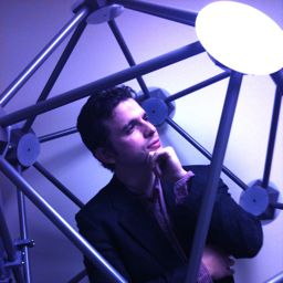
David Wozny, Ph.D.I am interested in how the human brain integrates streams of information within and across multiple sensory modalities. An even more critical feature is the ability to segregate and process information arising from multiple perceptual ‘objects’. Currently, I am investigating auditory processing and stream segmentation in background noise, and the associated perceptual limitations of cochlear implants. There is no question that the NITP program has substantially impacted my career. The program allowed for an easy transition to my first postdoc position at the Max Planck Institute for Human Cognitive and Brain Sciences in Leipzig Germany. The center is powerhouse of imaging modalities, and I was able to easily integrate myself into several research discussions surrounding a range of topics in EEG, MEG, DTI, TMS, tDCS, and fMRI. What impressed my peers the most is that in addition to having familiarity with each of these methodologies, I was also fully knowledgeable in the physics, hardware, and statistics that powered each of these tools. In my opinion, it is rare to find an imaging training program that could provide such a well rounded and structured learning environment. |
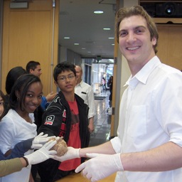 Dr. Wozny participating in Project Brainstorm |
|
I also know from my participation in the NITP summer school, that visiting students were also highly impressed with the program, and had many positive and encouraging words about the course. Naturally, such a program could not be possible without the dedication of the UCLA NITP faculty and staff. Present Position: Research Associate, Department of Otolaryngology, Oregon Hearing Research Center (OHRC), Oregon Health & Science University, Portland OR
|
||
 |
Rachel HigierI aim to work towards an empirically based model of psychopathology from a neurobiological perspective. I am interested in combining clinical information with neuroimaging and genetic data to investigate the diagnostic boundaries between Axis I disorders. I am particularly interested in how cognitive and affective impairments that are so often associated with varying psychiatric illnesses differ across and covary among specific disorders. My current work focuses on examining the genetic and neural factors that influence working memory function in patients with schizophrenia and bipolar disorder. During the NITP, I received excellent classroom instruction on a variety of neuroimaging methods from both a theoretical and practical perspective. Knowledge gained from this program helped inform my current research program aimed at integrating neuroimaging and genetic data to answer questions about etiological factors in severe mental illness. |
|
Present Position: Doctoral Student, Clinical Neuroscience Laboratory, UCLA |
||
Yan JinI am interested in using the advance MRI diffusion imaging tools, for example, High-angular resolution diffusion imaging (HARDI) to study human brain white tracts. Tractography can extract individual fibers. Once trajectories are extracted, we organize the curves into coherent bundles, by automated fiber clustering. I am currently researching on the automated fiber clustering algorithm over a large population study. Fibers must be clustered into recognizable bundles found consistently across subjects. NITP coursework is instrumental for the further development of my understanding in the neuroimaging research field. Especially, the two quarter series “Principles of Neuroimaging” will provide a comprehensive overview that will help me become familiar with other analytical methods and imaging modalities used in neuroimaging. The UCLA neuroimaging training program provides a unique interdisciplinary environment to anyone who has the passion in exploring the functions of our brain by utilizing various imaging modalities. |
 |
|
The most unique and signfiicant nature of this program for me is that it accepts international students, while most of the similar programs don't. I am an engineering student originally from China. I am currently working with Dr. Paul Thompson on developing automated white matter clustering algorithms with diffusion tensor images. The program provides indispensable and exhaustive knowledge on neuroimaging through a two-quarter basic training course and an advanced summer school. It has tremendously benefited my research so far. I also feel excited being in the program because I have an opportunity to talk to scholars from different research areas, not only limited to the field of engineering, which sometimes gives me surprising inspirations on my research. Moreover, the UCLA Advanced NeuroImaging Summer Program in 2010 was truly excellent in providing a basis for understanding the acquisition, processing, analysis, and interpretation of functional MRI data, which was obviously beneficial in my imaging research. I truly believe that NITP is establishing the solid foundation for me to being an aspiring neuroimaging researcher. Present Position: Graduate Student Research Assistant. Laboratory of Neuro Imaging (LONI) |
||
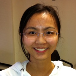 |
Xia HongjingMs. Xia is a biomedical engineer who is interested in the development of tools for neuroscientific research. Her current projects are in MR pulse sequences for rapid imaging, software development for real-time image processing, and critical evaluation and comparisons of signal quality in EEG systems. Hongjing is an international student from China. |
|
Present Position: Graduate student researcher, laboratory of Mark Cohen, UCLA
|
||
Leonardo MooreI am interested in integrative, multimodal approaches to studying social cognition, particularly:
|
 |
|
The educational and material resources provided by the NITP have allowed me to expand and improve on my existing research ideas as a function of my increased exposure to and in-depth comprehension ofneuroimaging. They have also served to deepen my understanding of thefundamental principles which underlie all of neuroimaging, which may one day allow me to design my own neuroimaging techniques to best suitmy projects (and not the other way around) Present Position: Graduate student researcher, laboratory of Susan Bookheimer, UCLA
|
||
 |
Angelica Morales, Ph.D.I am interested in using structural imaging techniques to characterize brain abnormalities in drug abusers and to see if there are changes in brain structure after prolonged abstinence or clinical intervention. When I began graduate school, I very little about neuroimaging methods. NITP helped me integrate what I knew about biology, mathematics and physics and apply it to image processing and experimental design. The foundation the course provided greatly facilitated my ability to learn and understand what was happening in the laboratory. Neuroimaging is a quickly evolving field, but the course's focus on fundamentals should make much of what I learned applicable in the future. |
|
Present Position: Graduate student researcher (T32 predoctoral fellow) The laboratory of molecular neuroimaging, UCLA
|
||
Nathan Hageman
|
 |
|
Hageman’s research focuses on diffusion tensor imaging (DTI), a type of magnetic resonance imaging (MRI) that allows visualization of the white matter tracts connecting different parts of the brain. He specializes in tractography – the use of computational methods to turn the data points acquired through DTI into a 3-D map of white matter tracts. Hageman has worked with established researchers at UCLA to apply these methods and investigate changes in brain connectivity associated with psychiatric and neurological disorders. For example, working with Katherine Narr, Ph.D., assistant professor of neurology at UCLA, he has reported that DTI is sensitive for detecting white matter atrophy in the temporal lobe of individuals with schizophrenia. Present Position: Graduate Student Research Assistant. Laboratory of Neuro Imaging (LONI) |
||
 |
Kathryn CrossI’m interested in automatic motor behaviors and their control. It is well-established that visuomotor transformations of various types can be automatic, and interference paradigms show that automatic motor activation from irrelevant stimulus features must be overcome to perform alternative actions. I am studying the mechanisms associated with overcoming these automatic motor activations, and am particularly interested in the stages of processing at which this control might occur and the potential for multiple mechanisms acting at different stages. The stages at which control of automatic motor behaviors occurs will also shed some light on whether there are distinct mechanisms for control over distinct visuomotor pathways, including those supporting spatial stimulus-response transformations, action observation and execution matching and object affordance. |
|
Present Position: Graduate student researcher, Laboratory of Dr. Marco Iacoboni, UCLA |
||
John ColbyIn general, my interests gravitate towards the intersection of science, engineering, and medicine,and the unique synergism that can result from their interaction. In line with this theme, my dissertation work includes:
The aims of the NITP closely align with those of my own career plan, and there is no doubt that participating in the NITP is one of the absolute highlights of my graduate education. |
 |
|
Present Position: MD/PhD student, Developmental Cognitive Neuroimaging Laboratory, UCLA
|
||
 |
April Ho, Ph.D.Dr. Ho is currently a Life Science Specialist with L.E.K. Consulting, an international strategy consulting firm that specializes in the life sciences industry. She joined L.E.K. in September of 2010 and has focused in the pharmaceutical (small and large molecule), genomics, and clinical diagnostics industries. |
|
Present Position: Life Science Specialist at LEK Consulting
|
||
N.B.: If you are an NITP fellow, please keep this page up to date by sending along comments, photos and news of your accomplishments.
The UCLA Neuroimaging Training Program is funded by generous awards from the National Institutes of Health, grant numbers R90 DA022768 and T90 DA023422